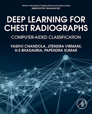
Deep Learning for Chest Radiographs: Computer-Aided Classification
暫譯: 胸部X光深度學習:電腦輔助分類
Chandola, Yashvi, Virmani, Jitendra, Bhadauria, H. S.
- 出版商: Academic Press
- 出版日期: 2021-07-22
- 售價: $3,760
- 貴賓價: 9.5 折 $3,572
- 語言: 英文
- 頁數: 228
- 裝訂: Quality Paper - also called trade paper
- ISBN: 0323901840
- ISBN-13: 9780323901840
-
相關分類:
DeepLearning
海外代購書籍(需單獨結帳)
商品描述
Deep Learning for Chest Radiographs enumerates different strategies implemented by the authors for designing an efficient convolution neural network-based computer-aided classification (CAC) system for binary classification of chest radiographs into Normal and Pneumonia. Pneumonia is an infectious disease mostly caused by a bacteria or a virus. The prime targets of this infectious disease are children below the age of 5 and adults above the age of 65, mostly due to their poor immunity and lower rates of recovery. Globally, pneumonia has prevalent footprints and kills more children as compared to any other immunity-based disease, causing up to 15% of child deaths per year, especially in developing countries. Out of all the available imaging modalities, such as computed tomography, radiography or X-ray, magnetic resonance imaging, ultrasound, and so on, chest radiographs are most widely used for differential diagnosis between Normal and Pneumonia. In the CAC system designs implemented in this book, a total of 200 chest radiograph images consisting of 100 Normal images and 100 Pneumonia images have been used. These chest radiographs are augmented using geometric transformations, such as rotation, translation, and flipping, to increase the size of the dataset for efficient training of the Convolutional Neural Networks (CNNs). A total of 12 experiments were conducted for the binary classification of chest radiographs into Normal and Pneumonia. It also includes in-depth implementation strategies of exhaustive experimentation carried out using transfer learning-based approaches with decision fusion, deep feature extraction, feature selection, feature dimensionality reduction, and machine learning-based classifiers for implementation of end-to-end CNN-based CAC system designs, lightweight CNN-based CAC system designs, and hybrid CAC system designs for chest radiographs.
This book is a valuable resource for academicians, researchers, clinicians, postgraduate and graduate students in medical imaging, CAC, computer-aided diagnosis, computer science and engineering, electrical and electronics engineering, biomedical engineering, bioinformatics, bioengineering, and professionals from the IT industry.
商品描述(中文翻譯)
《胸部放射線影像的深度學習》列舉了作者為設計一個高效的基於卷積神經網絡的電腦輔助分類(CAC)系統所實施的不同策略,該系統用於將胸部放射線影像進行二元分類,分為正常(Normal)和肺炎(Pneumonia)。肺炎是一種主要由細菌或病毒引起的傳染病。這種傳染病的主要目標是五歲以下的兒童和六十五歲以上的成年人,主要是因為他們的免疫力較差和恢復率較低。全球範圍內,肺炎的流行足跡廣泛,造成的兒童死亡人數超過任何其他免疫相關疾病,每年導致高達15%的兒童死亡,尤其是在發展中國家。在所有可用的影像學檢查方式中,如電腦斷層掃描(computed tomography)、放射線檢查(radiography或X光)、磁共振成像(magnetic resonance imaging)、超聲波(ultrasound)等,胸部放射線影像是最廣泛用於正常與肺炎之間的鑑別診斷的。這本書中實施的CAC系統設計使用了共計200張胸部放射線影像,其中包括100張正常影像和100張肺炎影像。這些胸部放射線影像通過幾何變換(如旋轉、平移和翻轉)進行增強,以增加數據集的大小,以便有效訓練卷積神經網絡(CNNs)。共進行了12次實驗,對胸部放射線影像進行正常與肺炎的二元分類。書中還包括了使用基於轉移學習的決策融合、深度特徵提取、特徵選擇、特徵維度減少和基於機器學習的分類器進行的深入實施策略,以實現端到端的基於CNN的CAC系統設計、輕量級的基於CNN的CAC系統設計以及混合CAC系統設計,專門針對胸部放射線影像。
本書是醫學影像、CAC、電腦輔助診斷、計算機科學與工程、電氣與電子工程、生物醫學工程、生物資訊學、生物工程以及IT行業專業人士的學者、研究人員、臨床醫生、研究生和本科生的重要資源。


![鐵路特考 2022 試題大補帖【電工機械大意(適用佐級)】(103~110年試題)(測驗題型)[適用電力工程]-cover](https://cf-assets2.tenlong.com.tw/products/images/000/170/855/medium/9786263270008.jpg?1638173413)


